
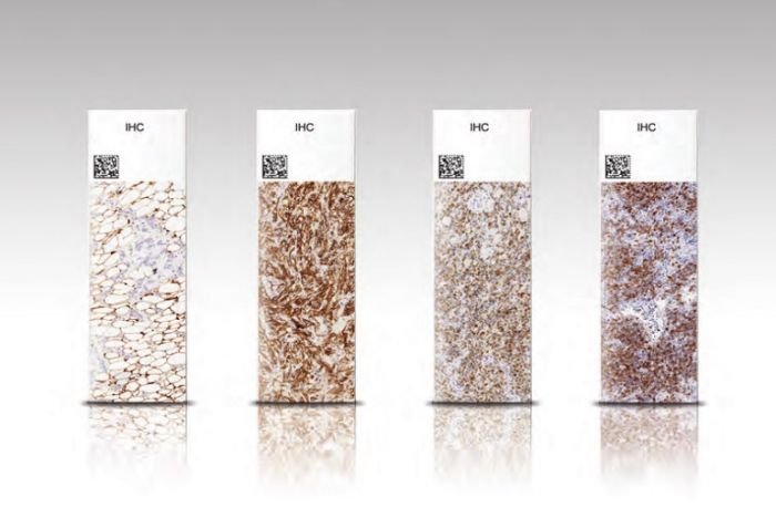
Immunohistochemistry (IHC), in situ hybridization (ISH) and other slide-based molecular tests are the more complex and clinically valuable tests performed in the histology laboratory. Advances in laboratory automation have made these technologies more accessible in the routine diagnostic workflow. However, staining quality and consistency is still variable within and between laboratories. Interestingly, the largest source of variability observed in an IHC stain is not due to the staining process, but due to the pre-staining processes as shown in Figure 1.

Standardization of fixation, grossing and tissue processing with advanced staining techniques are now widely recognized as critical in maintaining consistent, high quality stains. This is reflected in the 2013 ASCO/CAP guidelines for HER2 IHC testing in breast cancer, describing optimal conditions from fixation to staining. Standardization of these steps will only become more important with the growth of companion and molecular diagnostics as well as with the growth of ePathology all of which rely on consistency across the entire diagnostic process.
At Leica Biosystems our vison is to advance cancer diagnostics and improve lives. One way we can achieve this vision is by helping improve staining quality. As we recognize that IHC and ISH quality doesn‘t begin at the stainer, this series looks at many different aspects of staining quality, and considers how future tests will influence improved diagnosis.