Step 1 - Avoid Mechanical Trauma
 Tissue is removed gently to avoid trauma to the specimen caused by crushing or tearing. This applies both during surgery and during any further dissection that may be required of a fresh specimen.
 Specimen is damaged before fixation by crushing or tearing during removal.
Typical crush artifact is shown in this section of lymphoid tissue. It is characterized by dark, distorted cell nuclei, some of which are extremely elongated and intensely basophilic.
 |
Step 2 - Prevent Specimen Drying
Specimen is not allowed to dry out prior to fixation. If immediate fixation is not practicable, gauze moistened with saline can be used to prevent this.
Specimen is left on absorbent surface for some time prior to fixation.
This fresh specimen has just been removed from a patient during surgery. Because it is resting on an absorbent surface and the theatre is quite warm it will rapidly dry out unless it is immediately placed in fixative.
 |
Step 3 - Avoid Heat Damage
As far as possible avoid local heat damage to specimens (some damage by cautery may be unavoidable).
Any unnecessary local heat applied to tissue will cause damage. Fresh tissue is particularly susceptible.
A localized area at the edge of this breast specimen exhibits strong acidophilia with a loss of nuclear and cytoplasmic detail.
These effects are the result of heat damage caused when cautery was used during the removal of the specimen.
Adjacent glandular tissue is unaffected.
 |
Step 4 - Avoid Chemical Damage
Avoid contaminating fresh specimens with foreign chemicals or substances such as disinfectants.
The surface of unfixed tissue can be easily penetrated and damaged by foreign reagents or substances.
Monsel’s solution (ferric subsulphate solution) is a topical hemostatic agent used to control bleeding following mucosal biopsy. It causes coagulation and necrosis of the mucosal surface. If it is applied before a biopsy is taken it causes local basophilia and signs of early necrosis, masking pathological changes that may be present. Monsel’s solution artifact is most commonly seen when the patient is rebiopsied or wider excision is done later. The effects are seen in micrograph A of a H&E stained cervical biopsy. Micrograph B, stained with Perl’s method, shows the extensive deposition of iron on the specimen surface.
 |
Step 5 - Label Specimens Properly
Each specimen should be properly identified and all details recorded as soon as possible.
Recording of specimen details is delayed and the information provided is incomplete.
Specimens with incomplete labels such as these, should not be accepted by a laboratory. A procedure must be in place to deal with specimens that arrive at the lab inadequately labeled or accompanied by incomplete or inconsistent documentation.
 |
Step 6 - Ensure Prompt Fixation
Fixation is always carried out promptly. If it is necessary that a specimen remains unfixed for a short period of time, it should be refrigerated at 4 °C.
Fixation is delayed (degeneration of tissue elements commences as soon as the specimen is deprived of a blood supply).
A This autopsy liver specimen (H&E) shows the result of an extended delay before fixation. Note the poorly defined nuclei and imprecise cytoplasmic detail. Many bacteria are present within the central blood vessel.
B In this section of fibro-muscular tissue the nuclear chromatin is poorly preserved due to an extensive delay prior to fixation.
 |
Step 7 - Use Sufficient Fixative and a Suitable Container
An adequate volume of fixative (ratio of at least 20:1) is used in a container of an appropriate size. This avoids distortion of the fresh specimen and ensures good quality fixation.
Specimens are sometimes squashed into a small container with insufficient fixative to cover the specimen surface.
This container is too small for the mass of tissue it contains. There is insufficient fixative present and the specimen may well have been distorted as it was pushed into the container.
 |
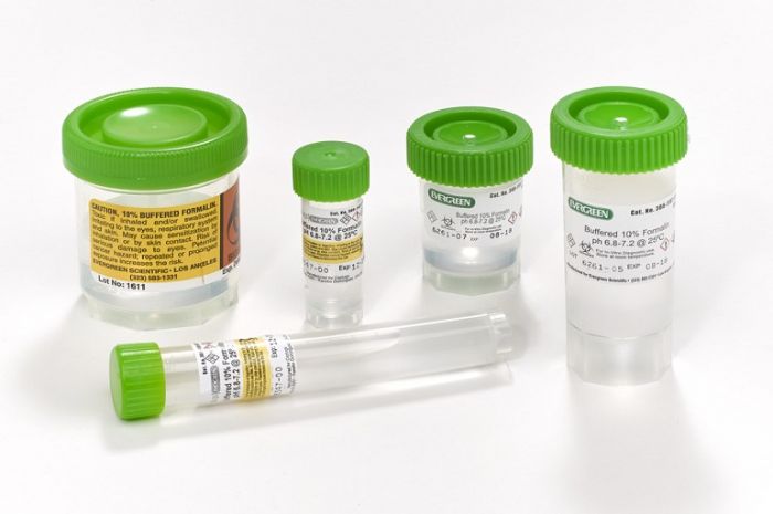
Step 8 - Check Fixative pH
The fixative is of high quality and at the optimal pH.
The fixative is of poor quality and unknown pH. If formalin is used at acid pH it rapidly produces “formalin pigment” by reaction with hemoglobin. Near neutral solutions will still produce the pigment but much more slowly. In good histological preparations formalin pigment should be removed prior to staining.
A The fixative used here has an unsatisfactory pH of 4.5. Buffered formalin solutions should have a pH of 6.8–7.0. B B The brownish-black granular deposit seen in the blood vessel in the center of this field is formalin pigment (acid formaldehyde hematin). It readily forms when tissue is fixed in acidic formalin and is usually seen in association with red blood cells.
 |
Step 9 - Expedite Large Specimen Fixation
The specimen dimensions allow rapid penetration of the fixative. Large specimens should be rapidly transported to the lab to allow grossing (tissue slices can be prepared to allow proper fixation to occur).
Large specimens are left in fixative for an extended time prior to grossing. The center of the specimen may remain unfixed and the tissue can become markedly distorted.
This large specimen (pig heart) has been sliced to allow the fixative access to all parts of the tissue. The slices are approximately 4–5 mm thick.
 |
Step 10 - Avoid Unnecessary Delays
No unnecessary delays – specimen reaches lab in minimum time.
Specimens are sometimes delayed – specimen transport has a low priority and is not well organized.
Priorities are important when delivering specimens to the laboratory. This is particularly so when frozen sections are involved.
 |
Step 11 - Handle Specimens Gently
Specimens handled gently – fragile specimens remain intact.
Specimens handled roughly – delicate friable specimens can be damaged.
Some of the tissue fragments seen at the bottom of this container have been produced by excessively rough handling during specimen transport. What was originally a cohesive specimen now consists of tissue fragments.
 |